Figure 2.
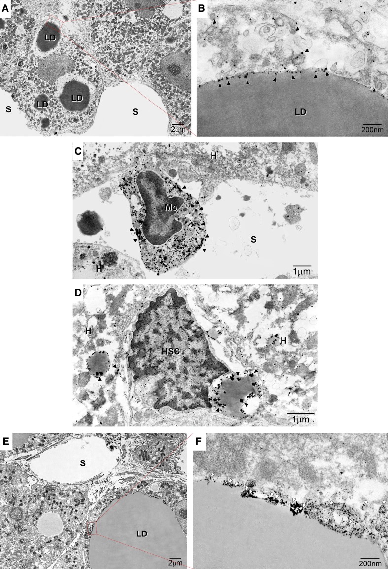
Immunoelectron microscopy of the subcellular localisation of glucagon-like peptide 1 receptor (GLP-1R) and caveolin-1 (CAV-1) in non-alcoholic steatohepatitis (NASH) liver. (A) Image showing marked macrovesicular steatosis: large lipid droplets (LDs) are visible in hepatocytes. (B) Immunogold particle labelling of GLP-1R on endoplasmic reticulum (ER)-like membranes attached to LDs and vesicles around LDs. (C) GLP-1R is localised to the peripheral rims of LDs in hepatic stellate cells and LDs in hepatocytes. (D) GLP-1R is localised to the plasma membrane and organelles of monocytes on hepatic sinusoids. (E) Macrovesicular and microvesicular LDs are visible in hepatocytes. (F) Immunogold particle labelling of CAV-1: CAV-1 is expressed on the caveolae of LDs. White arrowheads indicate CAV-1. A continuous basal lamina is apparent underneath the endothelium. Magnification=×20 000. Bar=200 nm. HSC, hepatic stellate cell. Arrowheads indicate CAV-1-positive particles. Black arrowheads indicate caveolae. Black arrows indicate LD caveolae. White arrows indicate ER-like membranes. Magnification=×2000. Bar=1 µm or 200 nm. White arrowheads indicate GLP-1R or CAV-1. Bar=200 nm. HSC, hepatic stellate cell. Black arrowheads indicate caveolae. Black arrows indicate LD caveolae. K denotes Kupffer cells. LD denotes lipid droplets.

