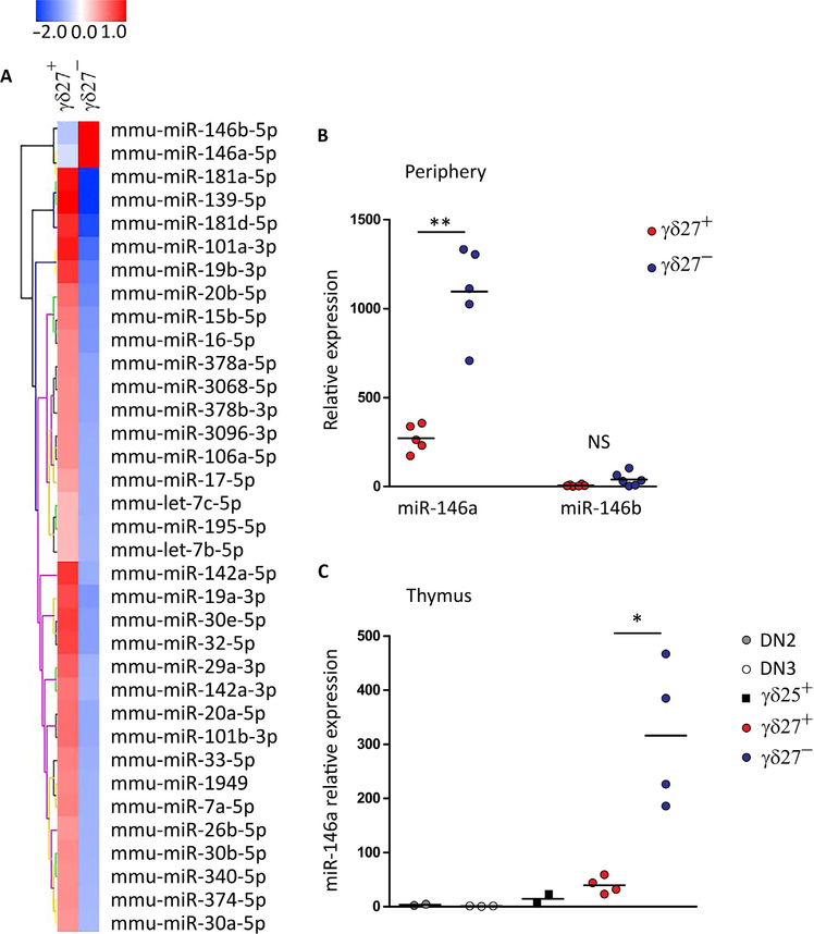
Fig. 1. miR-146a is highly expressed selectively on γδ27− T cells.
(A) Microarray heat map of differentially expressed miRNAs in duplicate samples of γδ27+ (n = 4 mice per sample) and γδ27−CCR6+ T cells (n = 8 mice per sample) isolated from pooled lymph nodes and spleen of C57BL/6 mice (more than twofold enrichment). (B) RT-qPCR analysis of miR-146a and miR-146b expression in sorted γδ27+ and γδ27− T cells from pooled peripheral organs (lymph node and spleen) of C57BL/6 mice. NS, not significant. (C) RT-qPCR analysis of miR-146a expression in sorted DN2 (CD4−CD8−CD44+CD25+), DN3 (CD4−CD8−CD44−CD25+), γδ25+ (CD25+CD27+), γδ27+, and γδ27− thymocytes of C57BL/6 mice. Results are presented relative to miR-423–3p or RNU5G (reference small RNA) expression. Each symbol in (B) and (C) represents an individual mouse. *P < 0.05 and **P < 0.01 (Mann-Whitney two-tailed test).

