
Figure 3.
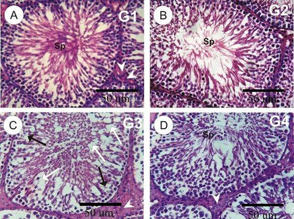
(A–D): photomicrographs of rat testes sections in the different experimental groups. A and B: testes sections in control and MLE rat groups showed normal seminiferous tubules lining spermatocytes and spermatogenesis. C: testis in tramadol group revealed the incomplete of spermatogenesis (black arrows), moderate degeneration (white arrows) in some seminiferous tubules with a decrease in the number of sperms, and depletion and little numbers of Leydig cells (arrowheads). D: testes in tramadol plus MLE showed the normal structure of seminiferous tubules with a normal distribution of the spermatogenic cells.