Figure 2.
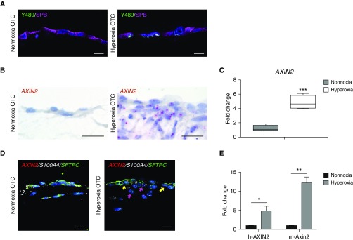
Hyperoxia exposure of three-dimensional organotypic cocultures (3D-OTC) stimulates active Wnt/β-catenin signaling. (A) Immunofluorescence for p-β-cateninY489 (nuclear β-catenin phosphorylated at tyrosine-489) (green), an established marker of Wnt activation, and SPB (surfactant protein-B) (purple) shows increased nuclear p-β-cateninY489 in hyperoxia-exposed OTC when compared with normoxic-cultured controls. Scale bars, 10 μm. (B) RNA in situ hybridization (RNA ISH) shows increased expression of Wnt target gene AXIN2 with hyperoxia exposure. Scale bars, 10 μm. (C) Real-time quantitative PCR (qPCR) comparing hyperoxia-exposed 3D-OTC with normoxia-cultured OTC shows increased expression of AXIN2. ***P < 0.01. (D) Multiplex RNA ISH showing increased expression of AXIN2 (red) in hyperoxia 3D-OTC relative to normoxia controls. AXIN2 is expressed in epithelial cells coexpressing SFTPC in green (yellow arrows) and mesenchymal cells expressing fibroblast marker S100A4 in white (magenta arrows). Scale bars, 10 μm. (E) MLE-15 cells were combined with human lung fibroblasts to make 3D-OTC, which were exposed to hyperoxia. Real-time quantitative PCR was performed with species-specific probes for AXIN2/Axin2. *P < 0.05 and **P < 0.01. Data are representative of three biological replicates for each condition, with three technical replicates for each sample used for qPCR.

