Figure 2.
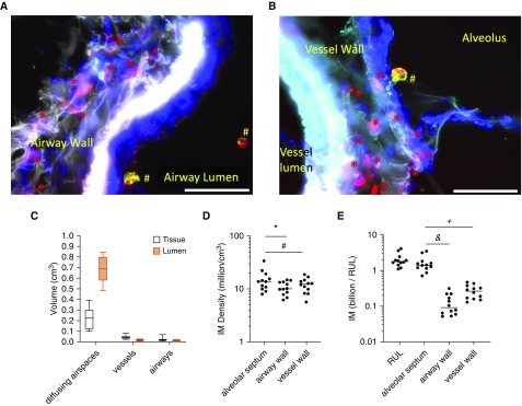
Quantification of interstitial macrophages (IMs) in lung subcompartments using stereology. (A) CD206 (cluster of differentiation 206)-positive IMs (red asterisks) are identified within the airway wall with CD206+/CD43+ airspace macrophages (red and green make yellow; #) located in the airway lumen. (B) Red CD206+ IMs visualized within the vessel wall and CD206+/CD43+ airspace macrophages (red and green make yellow; #) are visualized within the alveolus. Scale bars, 50 μm. (C) Volume fraction of each lung subcompartment was calculated via stereology. Diffusing airspaces consisted of alveoli, alveolar ducts, and respiratory bronchioles. (D) Density of IMs in lung subcompartments. *P = 0.02 and #P = 0.06. (E) Total number of IMs counted in human lung per subcompartment. &P < 0.0001 and +P < 0.003. RUL = right upper lobe.

