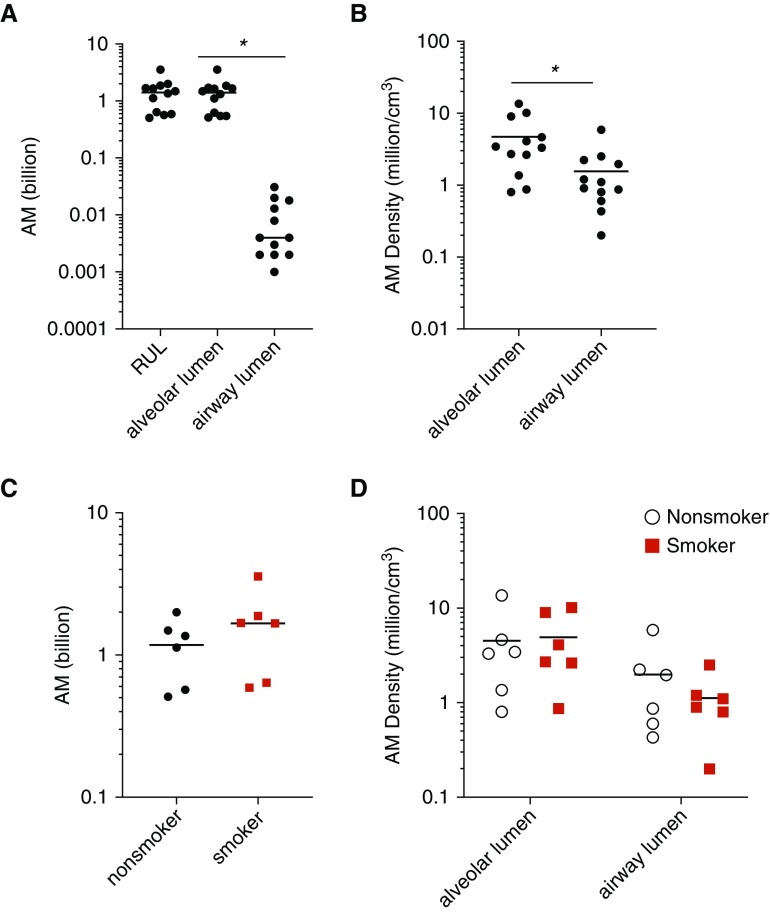
Figure 4.
Quantification of airspace macrophages (AMs) in human lung. (A) The overwhelming majority of AMs were present in the diffusing airspaces rather than the airway lumens. (B) The density of AMs in the diffusing airspaces was significantly greater than that within the airway lumens (by t test). (C) Total AM number was equivalent in nonsmokers and smokers. (D) AM density in tissue subcompartments was not different in smokers versus nonsmokers. Diffusing airspace was defined to include alveoli, alveolar ducts, and respiratory bronchioles. *P < 0.05. The y-axis is plotted as log10 scale. RUL = right upper lobe.

