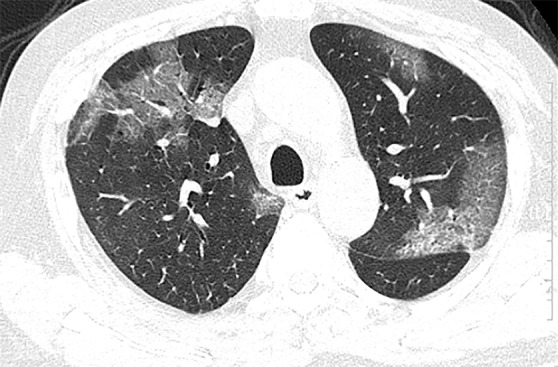
Figure 2a:

(a, b) Baseline CT images at admission of a 75-year-old man show multiple patchy areas of pure ground-glass opacity (GGO) and GGO with reticular and/or interlobular septal thickening. (c, d) Follow-up CT images on day 3 after admission show overlap of organizing pneumonia with diffuse alveolar damage in that it is more diffuse (not rounded) and associated with underlying reticulation, prominent progression with increased size and density of the lesions, and with more consolidations. Air bronchogram is also shown in d (arrows). Interlobular septal thickening does not seem to be major component. There is reticulation in many of cases of both suspected organizing pneumonia and diffuse alveolar damage.
