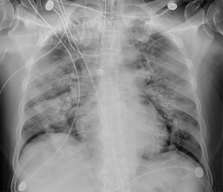
Figure 3d:

Serial imaging after admission of a 71-year-old man. (a) Baseline CT images on January 21, 2020, show consolidation of right upper lobe and ground-glass opacity (GGO) with consolidation and reticular and/or interlobular septal thickening of left upper lobe of left upper lobe; and patchy, focal, often rounded peribronchovascular and subpleural opacities associated with reticulation and architectural distortion. (b) Two days later, CT images show increased size of lesions in both lungs and decreased density in GGO lesions. (c) However, GGO on both lungs were larger on day 4 after admission. (d) Bedside portable chest radiograph on day 6 following admission shows diffusely increased opacities in both lungs, with relative bibasilar sparing.
