
Figure 4b:
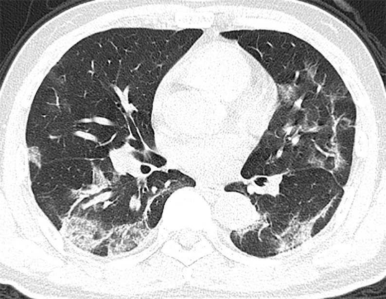
(a, b) Baseline CT images at admission of a 56-year-old man show multiple patchy areas of organizing pneumonia with some areas of interstitial and/or interlobular septal thickening and “strip shaped” consolidation (patchy, focal, often rounded, peribronchovascular and subpleural opacities associated with reticulation and architectural distortion). These abnormalities are mostly distributed in peripheral and posterior parts of lungs. (c, d) Follow-up CT images on day 5 after admission show interval improvement and absorption with fewer lesions and decreased lesion density.