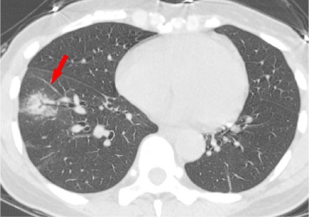
Figure 9a:

CT findings of confirmed Coronavirus Disease 2019 (COVID-19) pneumonia showing disease progression A 48-year-old woman presented with high fever (39.1 °C, 102.38℉) and Wuhan exposure history. a-b, On January 23, 2020, baseline axial unenhanced chest CT showed ground-glass opacity (GGO) with consolidation in lower lobe of right lung with typical air bronchogram (Panel a, arrow) and one pure GGO (Panel b, arrow) in the upper lobe of left lung. c-d, Three days later, follow-up axial unenhanced chest CT showed the disease progression, appearing as increased extent and consolidation (arrows) compared with baseline chest CT.
