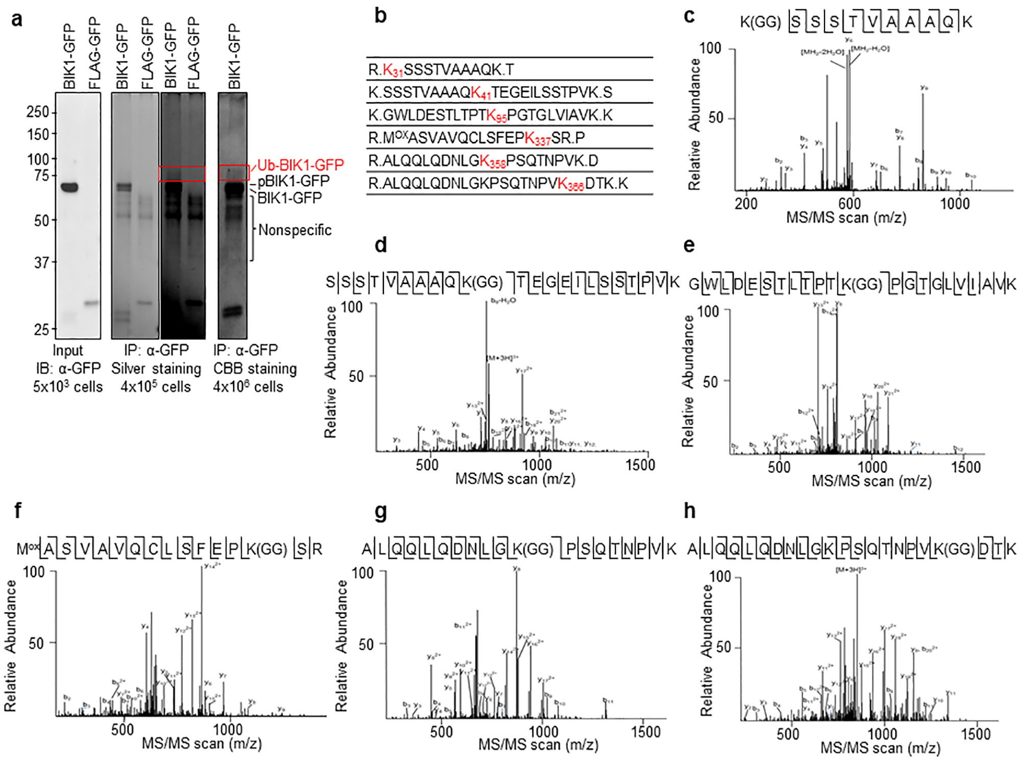
Extended Data Figure 7. BIK1 in vivo ubiquitination sites identified by mass spectrometry.

a. Ubiquitinated BIK1-GFP in planta was immunoprecipitated for LC-MS/MS analysis. BIK1-GFP and FLAG-UBQ were co-expressed in WT protoplasts (~4 × 10⁶ cells) followed by 200 nM flg22 treatment for 30 min. Ubiquitinated BIK1 was immunoprecipitated by GFP-trap-agarose, separated by SDS-PAGE, digested by trypsin and subjected to LC-MS/MS analysis. Portions of cell lysates were examined for BIK1-GFP expression (left), and immunoprecipitates were analyzed by SDS-PAGE following silver staining (middle, right for longer exposure of the same gel) and SDS-PAGE following CBB staining (right). The highlighted area was cut and analyzed by MS. b. BIK1 is ubiquitinated in vivo. Ubiquitinated lysine containing a diglycine remnant by LC-MS/MS analysis are marked as red with amino acid positions. c to h. MS/MS spectrums of peptides containing ubiquitinated lysine of BIK1 are shown. c. K31; d. K41; e. K95; f. K337; g. K358; h. K366. MS spectrums are outputs from the SEQUEST program. MS analysis was performed once.
