
Figure 1c:
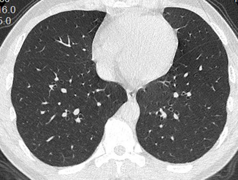
(a) Unenhanced CT image in a 43-year-old woman shows multiple ill-defined ground-glass opacities in the lateral segment of right lower lobe and the posterior segment of left lower lobe. (b) Unenhanced CT image in a 15-year-old male patient shows subtle nodular ground-glass opacities in the posterior segment of left lower lobe. (c) Unenhanced CT image in a 43-year-old man shows normal lungs.