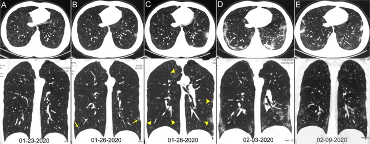
Figure 2:
Chest CT images of a 29-year-old man with fever for 6 days. RT-PCR assay for the SARS-CoV-2 using a swab sample was performed on February 5, 2020, with a positive result. (A column) Normal chest CT with axial and coronal planes was obtained at the onset. (B column) Chest CT with axial and coronal planes shows minimal ground-glass opacities in the bilateral lower lung lobes (yellow arrows). (C column) Chest CT with axial and coronal planes shows increased ground-glass opacities (yellow arrowheads). (D column) Chest CT with axial and coronal planes shows the progression of pneumonia with mixed ground-glass opacities and linear opacities in the subpleural area. (E column) Chest CT with axial and coronal planes shows the absorption of both ground-glass opacities and organizing pneumonia.

