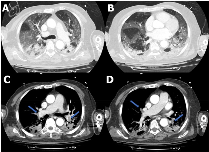
Figure 1.
Axial contrast-enhanced CT of the chest. A, B, Diffuse bilateral ground-glass opacities and small bibasilar consolidations compatible with COVID-19 pneumonia. C, D, Filling defects consistent with pulmonary emboli within the right upper lobe, right middle lobe, right lower lobe, and left lower lobe pulmonary arteries (arrows).

