
Figure 1c:
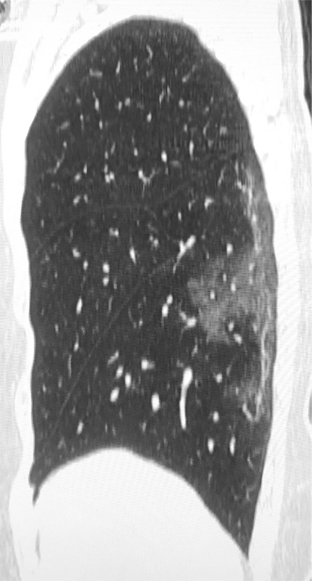
(a) Chest radiograph in a patient with COVID-19 infection demonstrates right infrahilar airspace opacities. (b) Axial and (c) sagittal chest CT images demonstrate peripheral right lower lobe ground-glass opacities. Follow-up (d) axial and (e) sagittal chest CT images 2 days later show improvement in the extent of ground-glass opacities, with more subpleural curvilinear lines (arrows).