
Figure 1e:
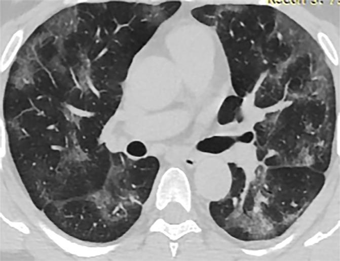
Chest CT findings in adult patients with COVID-19 on transaxial images. (a) Woman, 36 years old, 1 day after symptom onset. Subpleural ground-glass opacity in left lower lobe. (b) Man, 54 years old, 4 days after symptom onset. Subpleural ground-glass opacity in left lower lobe with inter- and intralobular septal thickening (crazy paving) and a ground-glass nodule in the right lower lobe (arrow). (c) Man, 28 years old, 3 days after symptom onset. Subpleural ground-glass opacity in the left lower lobe with central consolidation. (d) Woman, 49 years old, 7 days after symptom onset. Pure consolidation in right lower lobe. (e) Man, 42 years old, 6 days after symptom onset. Bilateral multifocal pure ground-glass opacities. (f) Man, 62 years old, 14 days after symptom onset, bilateral foci of consolidation in both lower lobes, with early linear opacities in a perilobular pattern (arrows).