
Figure 2c:
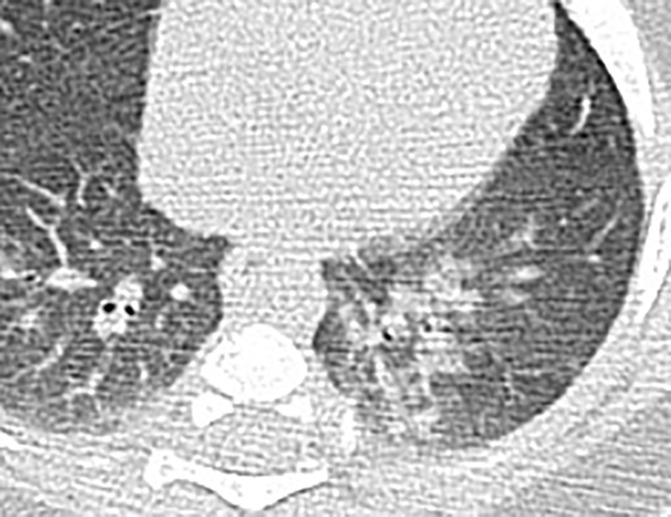
Chest CT findings of pediatric patients with COVID-19 on transaxial images. (a) Male patient, 2 months old, 2 days after symptom onset. Patchy ground-glass opacities (GGOs) in the right lower lobe. (b) Female patient, 4 years old, 4 days after symptom onset, two subpleural nodules in the right lower lobe. (c) Male patient, 8 months old, 6 days after symptom onset. Bronchial wall thickening and peribronchial GGOs and consolidation are noted in the left lower lobe.