
Figure 2a:
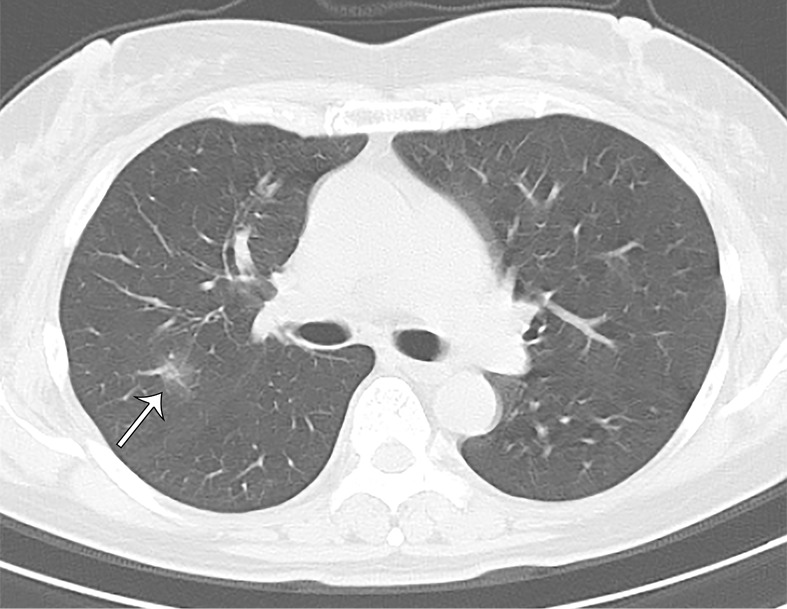
A 66-year-old asymptomatic woman. On axial CT images, focal rounded ground-glass opacities (arrows) with partial consolidation in a peribronchial and subpleural distribution were noted in the right upper (a), middle (b), and lower (c) lobes and left lower (b) lobe.