
Figure 3c:
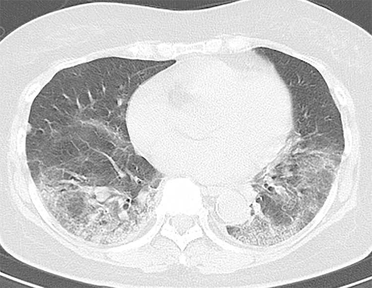
A 73-year-old asymptomatic woman. (a) On axial CT images, focal peripheral ground-glass opacities with intralobular and interlobular smooth septal thickening were shown in the left (arrowhead) and right upper lobe (arrows). The right upper lobe lesions were accompanied by subpleural curvilinear lines (arrow). (b, c) Diffuse ground-glass (reticular) opacities with consolidation with bronchiectasis and bronchial wall thickening were demonstrated in the left and right lower lobes.