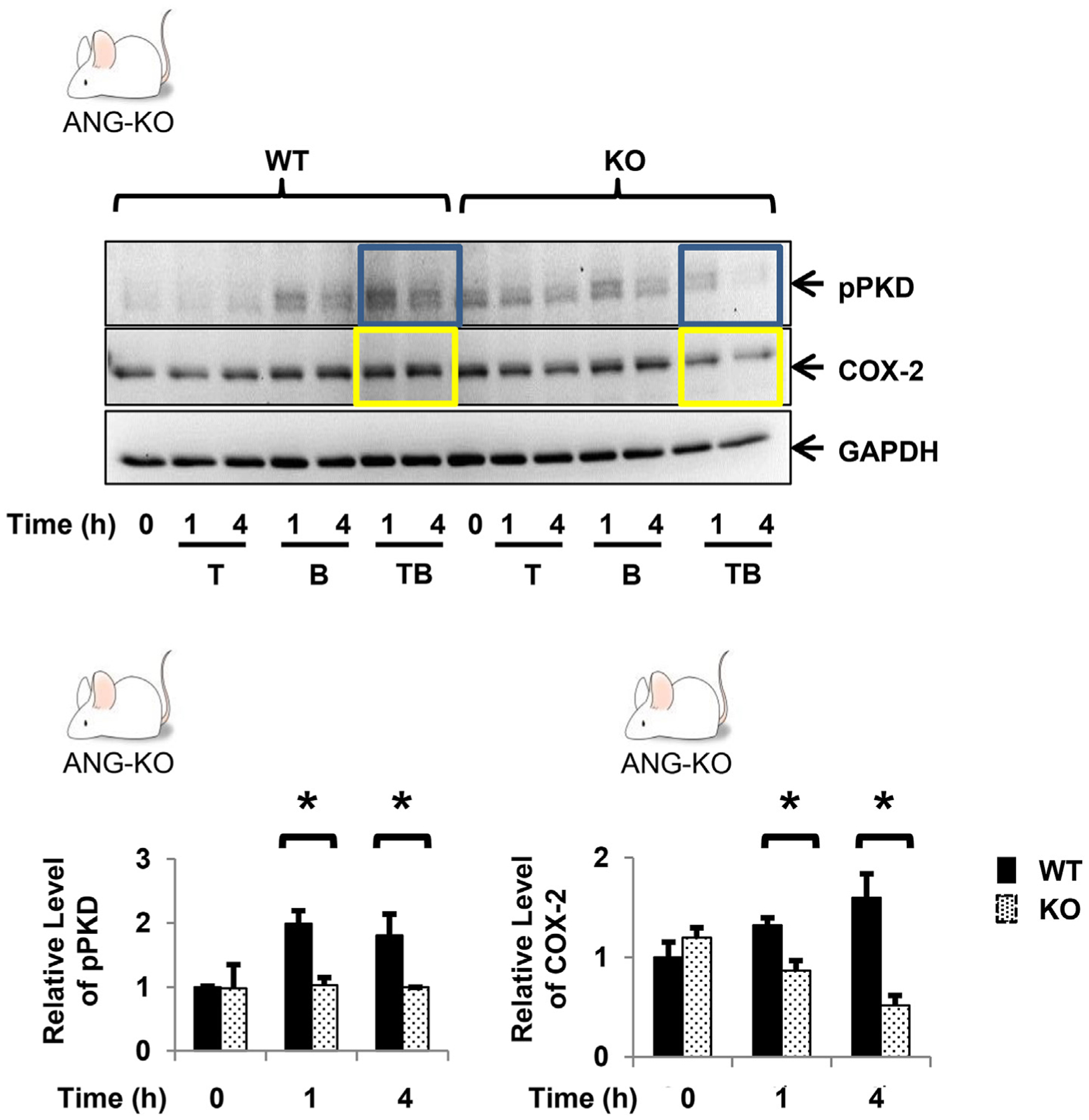
Fig. 4.

Confluent primary myofibroblasts from WT and ANG-KO mice were equilibrated in serum-free media for 30min and exposed to TNF-α (10 ng/ml) ± BK (100 nM) for up to 4h. The relative levels of pPKD and COX-2 following exposure to TNF-α (10 ng/ml) and BK (100 nM) are shown graphically, expressed as the mean ± S.E. n ≥ 3. * denotes p < 0.05.
