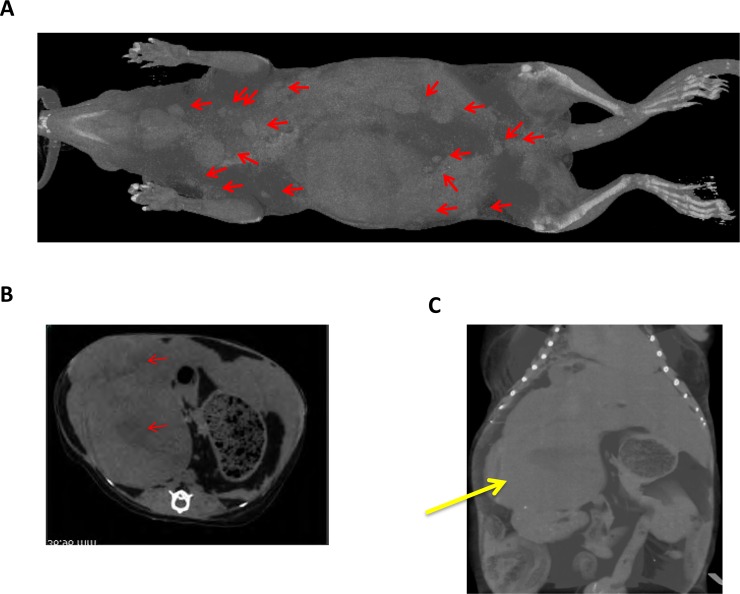
Fig 7. X-ray CT (LCT-200) analysis of a tumor-bearing tree shrew (No.26, 4 years 11 months old).
(A) Three-dimensional (3D) analysis of whole tree shrew body. Position of sarcoma was indicated by red arrows. (B) 3D tomogram of tree shrew. Arrows indicating tumor tissues. (C) 3D analysis of tree shrew belly. Yellow arrows indicate the large intraperitoneal tumors.

