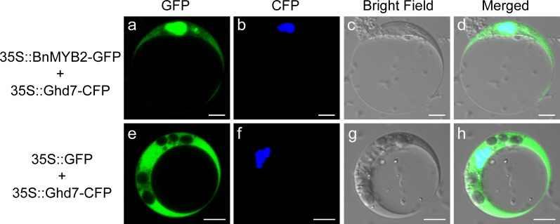
Fig 2. Subcellular localization of BnMYB2.
Constructs 35S::BnMYB2-GFP and empty vector 35S::GFP were separately co-transformed into Arabidopsis protoplasts with nuclear marker 35S::OsGhd7-CFP. GFP and CFP fluorescences were observed using a laser confocal microscope. a, 35S::BnMYB2-GFP; b, 35S::OsGhd7-CFP; c, Bright field; d, Overlap images of (a), (b) and (c); e, 35S::GFP; f, 35S::OsGhd7-CFP; g, Bright field; h, Overlap images of (e), (f) and (g). The bar indicates 5 μm.

