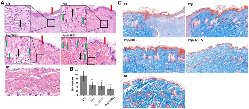
Figure 7.
Histological analysis of wound surface after treatment for 7 days. (A) H&E staining images. The red arrows mean newly formed skin surface. The black arrows mean infiltration of inflammatory cells. The green arrows mean neovascularization. (B) The thickness of newly formed skin surface. (C) Masson trichrome staining images

