Figure 2.

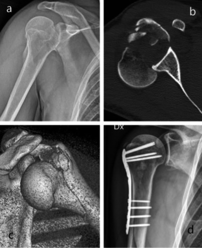
Imaging of patient G.M.: x-ray after trauma (a), axial ct of the fracture (b), 3d reconstruction of the fracture-dislocation (c), x-ray at follow-up (d)

Imaging of patient G.M.: x-ray after trauma (a), axial ct of the fracture (b), 3d reconstruction of the fracture-dislocation (c), x-ray at follow-up (d)