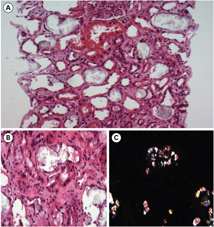
Fig. 2. Histopathologic evaluation. (A) Tubules reveal focal severe necrosis with crystal deposits and denudation of tubular epithelial cells. H&E stain (×20). (B) Aggregates of crystals attached to tubular basement membrane are shown with denudation of tubular epithelial cells. H&E stain (× 40). (C) The same image as Fig. 2B observed under the polarizing microscope.
H&E = haemotoxylin and eosin.

