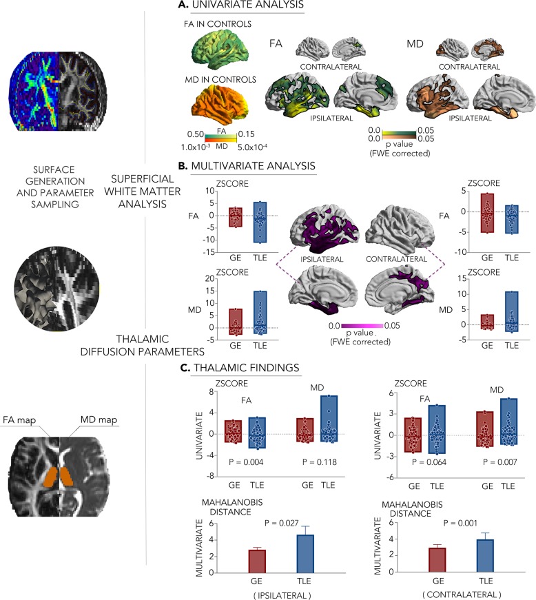
Fig. 2. Superficial white matter and thalamic diffusion MRI profiling (left).
a, b Uni- and multivariate superficial white matter differences between 107 patients with TLE and 96 with GE. Surface-based findings were corrected for multiple comparisons at a family-wise level of 0.05. c Uni- and multivariate findings in the thalamus. Line and edge of boxplots denote the mean and range of the value. The error bar represents standard deviation. For comparisons of each cohort relative to controls, see Supplementary Fig. 1b.

