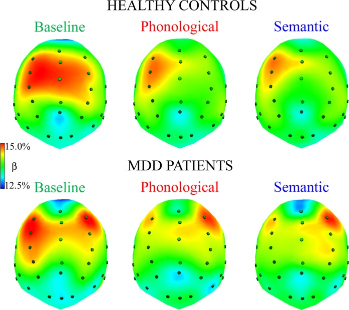
Figure 4.
Spline maps of normalized beta amplitudes during resting state, baseline condition (left panel), phonological and semantic linguistic tasks (middle and right panels, respectively) for HC and MDD patients. Qualitative analysis of spline maps suggests that, compared with controls, depressed patients had lower levels of beta rhythm in anterior regions of the left hemisphere during the two linguistic tasks. In addition, controls showed an anterior, left-lateralized pattern of activation (i.e., higher percentages of beta band in anterior left vs. right regions) and a bilateral posterior activation in all tasks.

