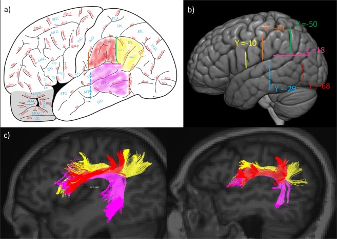
Figure 3.
(a) Delineation of the acceptable regions of interest (ROIs) for the dissected pathways. The angular gyrus (AnG in yellow) for SLF II, the supramarginal gyrus (SmG in red) for SLF III, and the posterior part of the temporal lobe (in fuchsia) on figure adapted from36, page 19, with permission. (b) Critical locations for ROI placement with coordinates in Montreal Neurological Institute (MNI) stereotaxic space: Yellow ROI (Y = −10), Orange ROI (Y = −21), Green ROI (Y = −50), Fuchsia ROI (Z = 18), Red ROI (Y = −68), Cyan ROI (Y = −29). The same Y and Z coordinates are valid for the right hemisphere. (c) Final dissection using DTI of the SLF II (in yellow), the SLF III (in red), and the AF (in fuchsia) for two participants.

