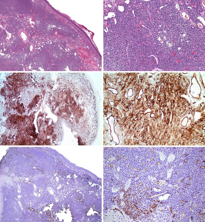
Fig. 1.
Glomangiopericytoma study case 4. (Top row, H&E) Spindled to epithelioid cellular proliferation with admixed staghorn-like vasculature with Grenz zone underlying sinonasal mucosa. (Middle row; CD34 from Leica) Diffuse moderate reactivity for CD34 in lesional cells. (Bottom row; CD34 from Ventana) Patchy weak reactivity for CD34 in lesional cells

