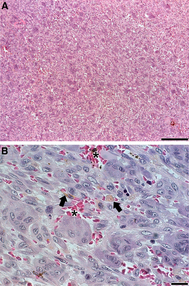
Fig. 5.

a, b Incisional biopsy shows characteristic histopathologic features of central giant cell granuloma. Multinucleated giant cells are separated by interstitial fibrous connective tissue bands. Hemosiderin pigment (arrows) and areas of haemorrhage (asterisks) were present (stained with H&E). Scale bars: a, 200 µm and b, 20 µm
