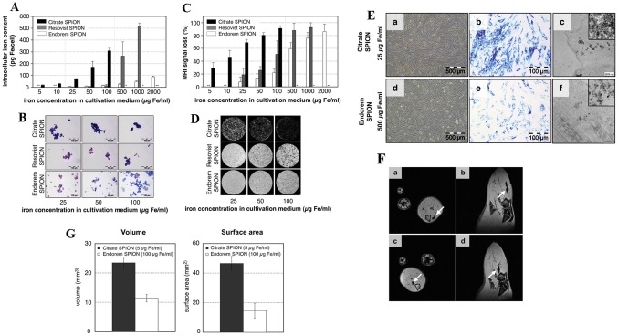
Fig. 1.
Analysis of the intracellular iron content in cultivation medium using a ICP–OES, prussian blue staining b, and visualization with MRI in vitro c, d. e Intracellular Imaging of SPIONs after incubation with Citrate and Endoderm SPION using microscope, Prussian blue staining, and TEM. f In vivo MR imaging of Citrate SPION (a, b) and Endoderm SPION (c, d) labelled cells which showed hypointensity in rat muscle tissue in the axial and sagittal.
(Reprinted with from Andreas et al. [47]. Copyright (2012), with permission from Elsevier)

