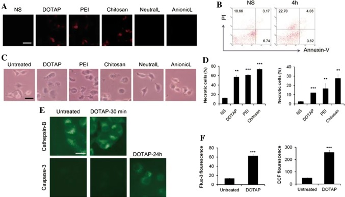
Fig. 5.
a Analysis of necrosis by propidium iodide in mouse lungs, b flow cytometry of Annexin-V and PI to detect necrotic cells induced by injecting cationic liposomes, c microscopic images of cell morphological changes induced various nanocarriers in vitro. d Quantification of flow cytometry to detect necrotic cells using PI and Annexin-V after nanocarriers treatment. e Immunofluorescence image of Cathepsin-B and Caspase-3 after treatment of DOTAP with time intervals. f ROS levels and Ca2+ concentration detected by H2DCF-DA and Fluo-3/AM with flow cytometry in A549 cells after DOTAP liposomes treatment.
(Reprinted with from Wei et al. [87]

