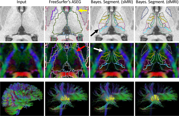
Fig. 3.
Top two rows: axial slice of a T1 scan and principal diffusion directions of an HCP subject, with segmentations from FreeSurfer, Bayesian segmentation (T1 only), and the proposed method (T1+dMRI). Bottom row (left to right): Whole-brain tractography (25,000 tracts); subset of tracts going through the thalami (in yellow) as segmented by: FreeSurfer (2,602 tracts); Bayesian segmentation of T1 (2,193 tracts); and proposed method (1,676 tracts). See Section 3.3 for a description of the arrows.

