Fig. 4.
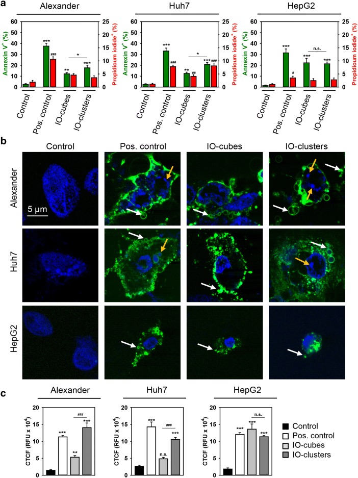
Analysis of apoptotic cell death upon treatment with IO-cubes and IO-clusters. a Cells were stimulated with IO-cubes or IO-clusters (100 µg/mL) for 24 h and labeled with annexin V—green dye, propidium iodide—red dye, hoechst 33342 nuclear stain—blue. Labeled cells were imaged with epi-fluorescence microscopy. ImageJ software (NIH) was used for calculation of annexin V and propidium iodide positive cells. Data are expressed as mean ± SEM (n = 3), *p < 0.05, **p < 0.01, ***p < 0.001, #p < 0.05, ##p < 0.01, ###p < 0.001. Cells treated with 2 μM staurosporine for 3 h served as a positive control. b Cells were stimulated with IO-cubes or IO-clusters (100 µg/mL) for 24 h and then labeled with hoechst 33342 nuclear stain (blue) and annexin V (green). Yellow arrows indicate nuclear fragmentation; white arrows – blebbing of cytoplasmic membranes. Cells treated with 2 μM staurosporine for 3 h served as a positive control. Labeled cells were then imaged using high-resolution spinning disk confocal microscopy (Spin SR, Olympus). c Caspase-3 activation in hepatic cell lines. Alexander, HepG2 and Huh7 cells were stimulated with IO-cubes or IO-clusters (100 µg/mL) for 24 h, and incubated with fluorescein-conjugated pan-caspase inhibitor (VAD-FMK). Following the staining, cells were analyzed using a spinning disk confocal microscopy. Quantification of fluorescence intensities was performed in ImageJ (NIH) software. Data are expressed as mean ± SEM (n = 3), **p < 0.01, ***p < 0.001, ##p < 0.01, ###p < 0.001. Cells treated with 2 μM staurosporine for 3 h served as a positive control

