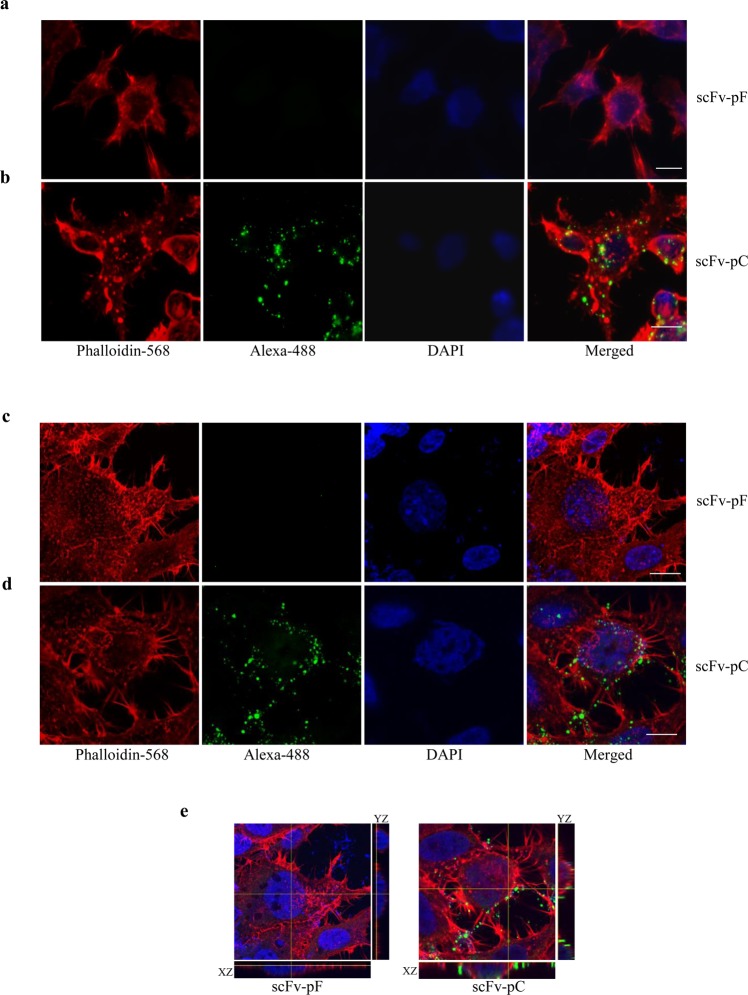
Figure 3.
Intracellular delivery of scFv by CPP in SH-SY5Y and HEK293T cells. SH-SY5Y (a,b) or HEK293T (c,d,e) cells incubated with scFv-pF and -pC for 120 min in Opti-MEM were fixed and stained using anti-His antibody and Phalloidin-594 and imaged using confocal microscope. Serial optical sections in the Z-axis of the cell, collected at 1 μm intervals with 63x oil immersion objective lense (NA 1.4) were projected and observed in a total thickness of 10 μm by using LSM 780 (version 3.2) software. scFv-pF (a or c) is not showing any specific staining indicating no intracellular delivery due to the lack of any cell penetrating peptide. scFv-pC (b,d) is showing good specific staining around the cells and inside cells indicating its binding to the cell membrane and possibly intracellular delivery due to the presence of CPP-peptide. (e) Merged confocal image with the orthogonal projection of scFv-pF (left) and scFv-pC (right) localization along with XZ and YZ views. Scale bars = 10 μm.

