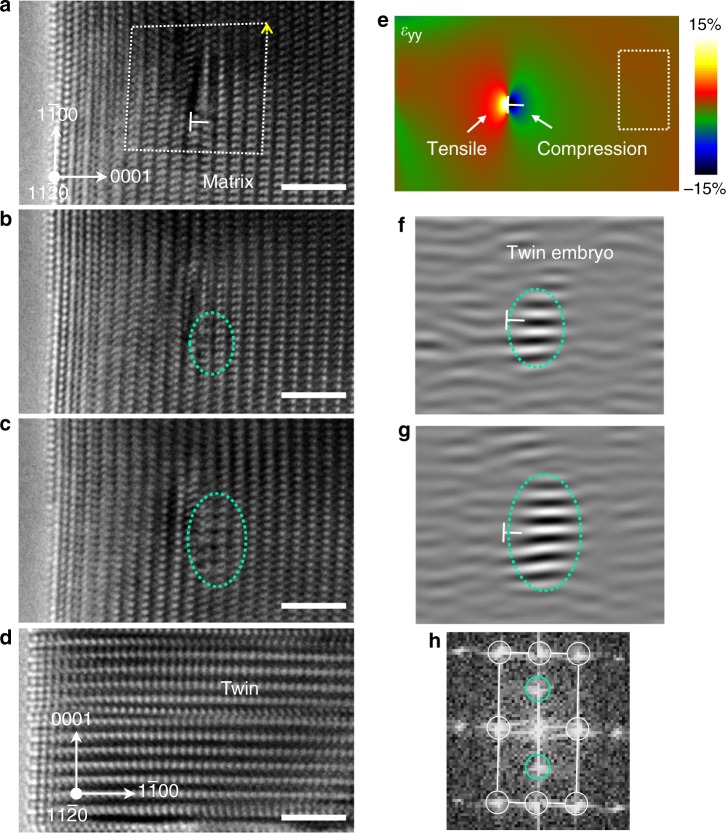
Fig. 5. Twinning nucleation assisted by the strain field of a matrix dislocation.
a–d Sequential HRTEM images showing the nucleation of a twin embryo near the dislocation core (indicated by the “├” symbol). The crystal was under <1 −1 0 0 > -oriented compression and viewed along the <1 1 −2 0> direction. White dashed line in a is the Burgers circuit which indicates an <a > component in the Burgers vector (indicated by the yellow arrow) of the dislocation. Detailed analysis of the Burgers vector is shown in Supplementary Fig. 5. e Mapping of the strain normal to the parent prismatic plane (εyy) generated by the dislocation core, by geometrical phase analysis. Boxed region is the reference region for the geometrical phase analysis (see ref. 56 for more details). f–g Inverse fast Fourier transformations of the TEM snapshots in b, c, respectively, using the (0 0 0 1) reflections of the twin (indicated by turquoise-circles in h). Turquoise dotted circles in b, c, f, g mark the twin embryo. White circles and lines in h indicate the reciprocal lattice of the matrix. Scale bars in a–d, 2 nm.

