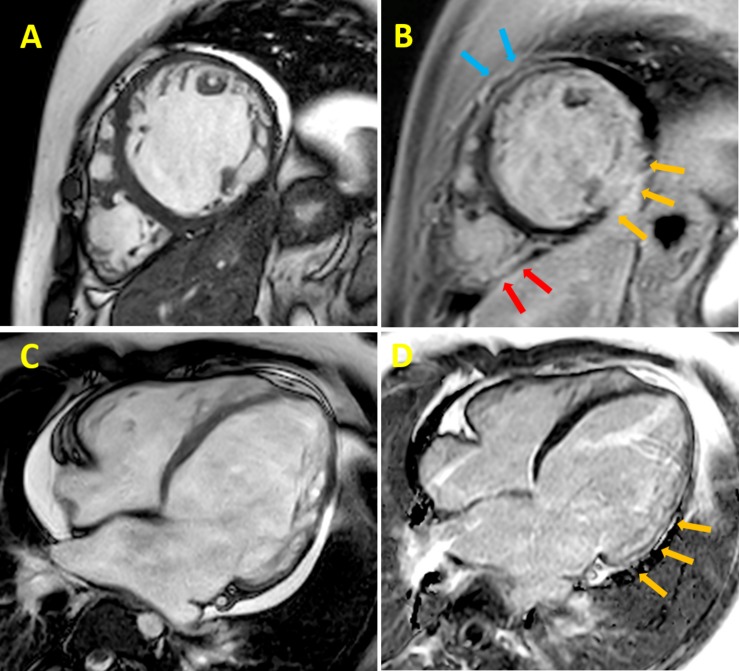
FIGURE 1.
Cardiac magnetic resonance (CMR) cine imaging of a 24 years old DMD patient. The collection of representative images from patients was approved by the Ethical Committee of the Meyer Pediatric Hospital of Florence in the context of a project funded by Telethon Italy (grant GGP16191). Informed consent to patients was performed conform the declaration of Helsinki. This data does not contribute to any novel finding. (A) Late gadolinium-enhancement (B) left ventricular (LV) short-axis section images of a patient with Duchenne muscular dystrophy (DMD). Yellow arrows indicate the inferolateral subepicardial and midwall contiguous fibrosis; light blue arrows indicate the anterior segment and the red arrows the posterolateral right ventricle wall, both showing midwall fibrosis; CMR cine imaging (C) and late gadolinium-enhancement (D) LV long-axis section images of the same patient with DMD; yellow arrows indicate midwall fibrosis of the inferolateral segment.

