Figure 1.
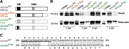
Description of the zebrafish models used. A, Schematic structure of the predicted Dmrt3a protein, showing the DNA binding domain (DM) and the Dmrt3 family domain (DMA). Lighter shade represents missing amino acids compared with the wild-type form of Dmrt3a. B, Western blot for Dmrt3a (47 kDa) and β-actin (42 kDa) protein at 3 dpf in dmrt3aWT, dmrt3a47aa, and dmrt3a376aa as well as 2 and 3 dpf in dmrt3aMO. C, Alignment of dmrt3a cDNA partial sequences between dmrt3aWT (top) and the CRISPR/Cas9 generated mutant dmrt3a47aa(bottom). The fragment shows the proximity to the 5 bp deletion (—) where the asterisk (*) represents the premature stop codon generated and the underlined sequence indicates the sgRNA target. This figure is extended in Extended Data Figure 1-1.

