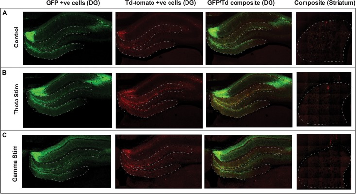
FIGURE 3.
Confocal immunofluorescence imaging of representative hippocampal dentate gyrus and striatum adjacent to the virus injection site. Native fluorescence from the repeat stimulation experiment is shown in the three stimulation groups: (A) Control (no stimulation) (B) Theta stimulation (C) Gamma frequency stimulation. The green GFP channel (green) serves as a reporter for transduced hippocampal neurons, whereas the Td Tomato channel (red) shows the transduced cells with concomitant c-Fos expression.

