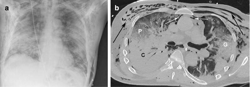
Fig. 1.
a. Chest X-ray demonstrating diffuse subcutaneous emphysema and bilateral pulmonary infiltrates. b. High-resolution chest CT showing an association of consolidations (C), ground glass opacities (G) and crazy paving pattern (P) highly suggestive of COVID-19. Pneumomediastinum is well identified (white arrows) as well as subcutaneous emphysema (black arrows)

