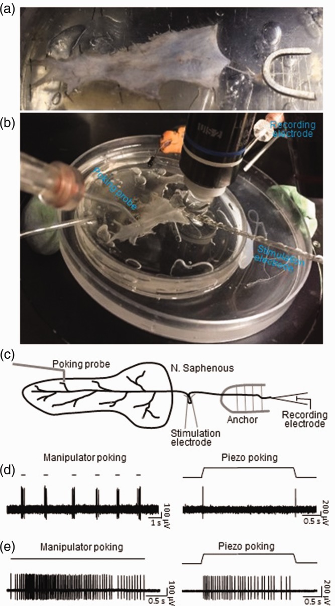
Figure 2.
Application of the pressure-clamped single-fiber recordings to study mechanical receptors in skin-nerve preparations of mice. (a) Image showing a skin-nerve preparation made from a piece of shaved hairy skin from a mouse hindpaw and the attached saphenous nerve. The skin was affixed to the bottom of the recording chamber by tissue pins and the saphenous nerve was affixed with a tissue anchor. An actual image (b) and schematic diagram (c) of experimental settings on the stage of an upright microscope during the pressure-clamped single-fiber recordings from the skin-nerve preparation. (d) Sample traces showing impulses recorded from a single saphenous nerve fiber following mechanical probing manually operated by a manipulator (left, manipulator probing) or by a computer-programmable piezo device (right, piezo probing). Manipulator probing was applied briefly for multiple times with the indentation step of 100 µm (left). Piezo probing was applied in a ramp-and-hold indentation step of 38 µm for a duration of 2.5 s (right). The mechanoreceptor of this recording shows to be a typical RA type. Similar results were obtained in seven other single-fiber recordings. (e) Sample traces showing impulses recorded from another single saphenous nerve fiber following manipulator probing (left) or piezo probing (right). Manipulator probing was applied with indentation step of 100 µm for a prolonged period of 3.2 s (left). Piezo probing was applied in a ramp-and-hold step of 38 µm and for a duration of 2.5 s (right). The mechanoreceptor of this recording shows to be a typical SA1 type. Similar results were obtained in two other single-fiber recordings.

