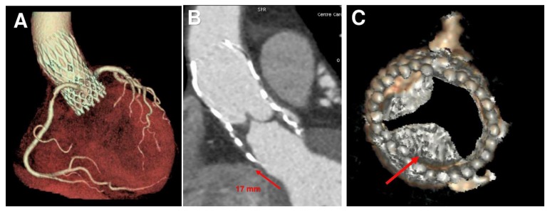
Figure 1.
Transcatheter aortic valve replacement (TAVR) position, thrombus formation and biomechanical model of patient-specific aortic root from medical CT images. (A) Pre-operative CT scan of CoreValve (26 mm). (B) Post-operative CT scan at one year shows a device upward shift. (C) Thrombus formation in subvalvular zone (red arrow).

