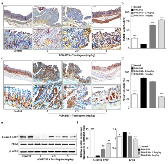
Figure 4.

Tussilagone treatment induces apoptosis and inhibits cell proliferation in the colon. (a) Apoptosis was measured via terminal deoxynucleotidyl transferase dUTP nick end labeling (TUNEL) staining of the colon sections of mice (upper, ×100 magnification; lower, ×400 magnification; bar = 100 µm). (b) The percentage of TUNEL-positive cells in the colon tissues was estimated as described in Methods. (c) The cell proliferation was measured via PCNA staining of the colon sections from mice (upper, ×100 magnification; lower, ×400 magnification; bar = 100 µm). (d) The percentage of PCNA-positive cells in the colon tissues was calculated as described in Methods. (e) The protein samples of the whole-cell lysates from the colon tissues were separated by SDS-PAGE. Cleaved-PARP and PCNA were detected by Western blot and visualized by chemiluminescence. Photos are representative images. (f) The quantitative data were normalized by internal control, β-actin and further expressed as folds, presented as the comparison with the amount relative to the AOM/DSS-treated mice. The results are expressed as the mean ± SEM, * P < 0.05, ** P < 0.01, *** P < 0.001, compared with the AOM/DSS-treated mice.
