Figure 5.

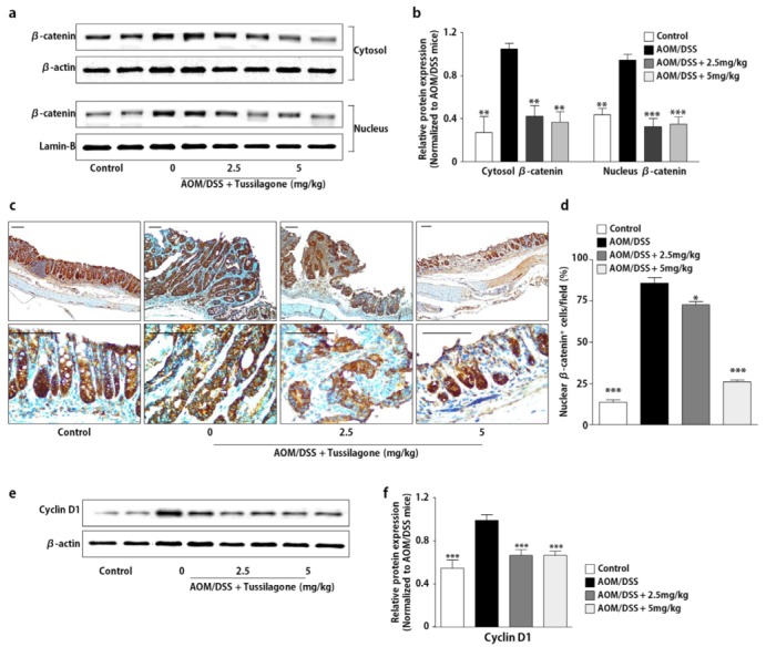
Administration of tussilagone inhibits the β-catenin expression in the colon tissues. (a) The protein samples in the cytosol or nuclear fraction from the colon tissues were separated by SDS-PAGE. β-catenin was detected by Western blot and visualized by chemiluminescence. Photos are representative images. (b) The quantitative data were normalized by internal control (β-actin for cytosol; lamin-B for nuclear fraction) and further expressed as folds, presented as the comparison with the amount relative to the AOM/DSS-treated mice. (c) The immunohistochemical analysis of nuclear β-catenin expression in the colon tissues (upper, ×100 magnification; lower, ×400 magnification; bar = 100 µm). (d) The percentage of nuclear β-catenin positive cells in the colon tissues was calculated as described in Methods. (e) The protein samples of the whole-cell lysates from the colon tissues were separated by SDS-PAGE. Cyclin D1 was detected by Western blot and visualized by chemiluminescence. Photos are representative images. (f) The quantitative data were normalized by internal control, β-actin and further expressed as folds, presented as the comparison with the amount relative to the AOM/DSS-treated mice. The results are expressed as the mean ± SEM, * P < 0.05, ** P < 0.01, *** P < 0.001, compared with the AOM/DSS-treated mice.
