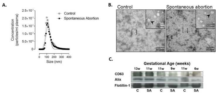
Figure 1.
Characterization of extracellular vesicles (EVs) isolated from plasma using ExoQuickTM in controls and patients who had spontaneous abortion. (A) Mean particle diameter of EVs obtained from plasma for all the samples by nanoparticle tracking analysis. (B) Representative image of transmission electron microscopy of the EVs obtained. [ ], representes area zoomed in the upper right corner with 100nm; arrows indicate extracellular vesicles. (C) Protein extracted from EVs was isolated and analyzed by Western blot at different gestational ages to determine the protein expression levels of CD63, Alix, and Flotillin-1.

