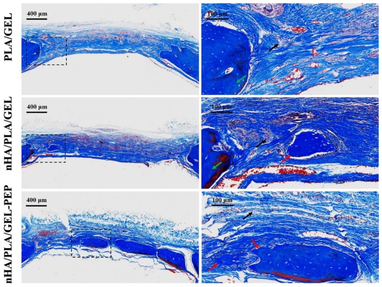
Figure 4.
Masson’s trichrome-stained images of the newly formed bone within the repaired tissue eight weeks after surgery, using scaffolds made of PLA/gelatin (GEL), nano-hydroxyapatite (nHA)/PLA/GEL and nHA/PLA/GEL/BMP-2 peptide (-PEP). Red arrows indicate new bone, green arrows indicate host bone and black arrows indicate residual scaffolds. Residual scaffolds were all clearly filled with intercellular collagen fibers stained blue, and the newly formed bone tissue was dark blue because of the existence of abundant and compact collagen. New bone regenerated in the nHA/PLA/GEL-PEP group existed both in the middle and limbic of the defects (used with permission from [171]).

