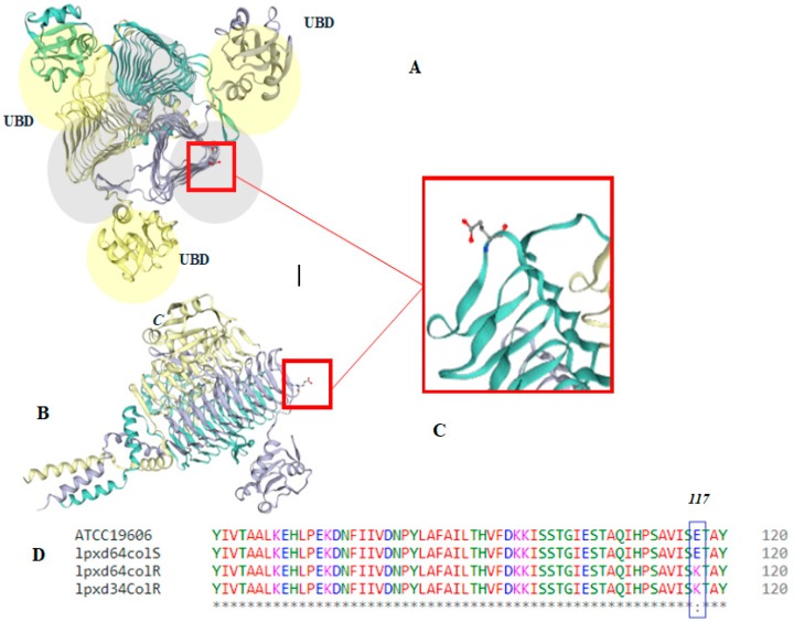
Figure 2.
Predicted 3D model structure of MS64Col-R LpxD protein. (A) Orthogonal view of the trimer, showing three chains where the lipid-binding domain (LBD) is colored in grey shades. (B) Side view of the trimer. (C) Amino acid substitution position (117). (D) Amino acid sequence alignments of a part of the LpxD protein of the colistin-sensitive reference strain (ATCC19606), colistin-sensitive clinical isolate (64colR), colistin-resistant laboratory-induced isolate (64colR), and colistin-resistant clinical isolate (34colR), generated by online Clustal W–Model server. UBD: uridine-binding domain.

