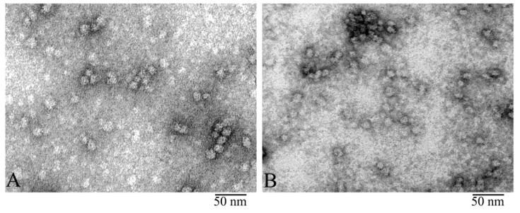
Figure 1.
Electron microscopy (EM) of negatively stained α-crystallin from bovine eye lens: Comparison of natural α-crystallins (A) and the preparation produced by Sigma (B). EM images show that both preparations form heterogeneous complexes identical in shape and dimensions. Preparation conditions for EM analysis: C = 0.2 mg/ ml, incubation for 30 min at 37 °C in 20 mM Tris-HCl buffer (pH 7.5), 100 mM NaCl, and 1 mM EDTA.

