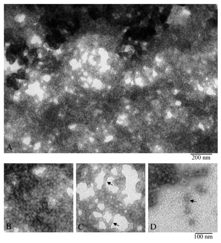
Figure 6.
EM images of α-crystallin gel (nucleus). Fragments of the fields of α-crystallin gel: (A,B) field fragments with tight packing of heterogeneous oligomer complexes; (C) field fragment with lower density of α-crystallin complexes, arrows show short stacks of complexes; and (D) field fragment with single complexes (shown by the arrow).

