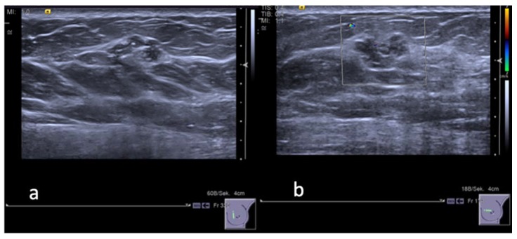
Figure 2.
A 48 years old woman. (a) Gray-scale ultrasound image shows a hypoechoic «mass» lesion with lobulated and partially non circumscribed margins in the inner para-areolar region of the left breast. The maximum diameter of the lesion was 14 mm. (b) Color Doppler image reveals no peripheral vascularity of the lesion. The lesion was categorized by both readers as BI-RADS 3. CNB: B3 lesion without atypia (adenosis, sclerosing adenosis, epithelial proliferation without atypia, small papilloma). Final histology after surgery revealed a highly differentiated, intraductal carcinoma DCIS-G1, in addition to extensive adenosis, small papilloma, and lobular neoplasia.

